Your heart beats roughly 100,000 times a day, and each beat moves blood through two separate loops: one that visits the lungs and one that serves the rest of the body. It is easy to picture the heart as a single pump that pushes blood around in one circle. The real arrangement is more interesting, and understanding it makes sense of terms you hear in everyday health conversations, from "blood pressure" to "heart valves" to "heartbeat."
This article is an educational overview of normal heart physiology. It is not medical advice and cannot be used to diagnose or treat any condition. If you have concerns about your own heart, speak to a qualified clinician.
Key takeaways
- The heart is a muscular organ with four chambers: two upper atria and two lower ventricles.
- It runs two circuits in series. The right side sends blood to the lungs; the left side sends blood to the body.
- One-way valves keep blood moving forward. They open and close in response to pressure differences, not because they are "controlled."
- A small cluster of cells called the sinoatrial (SA) node sets the normal rhythm using electrical signals.
- Arteries carry blood away from the heart and veins carry it toward the heart. That definition does not depend on how much oxygen the blood holds.
The four chambers
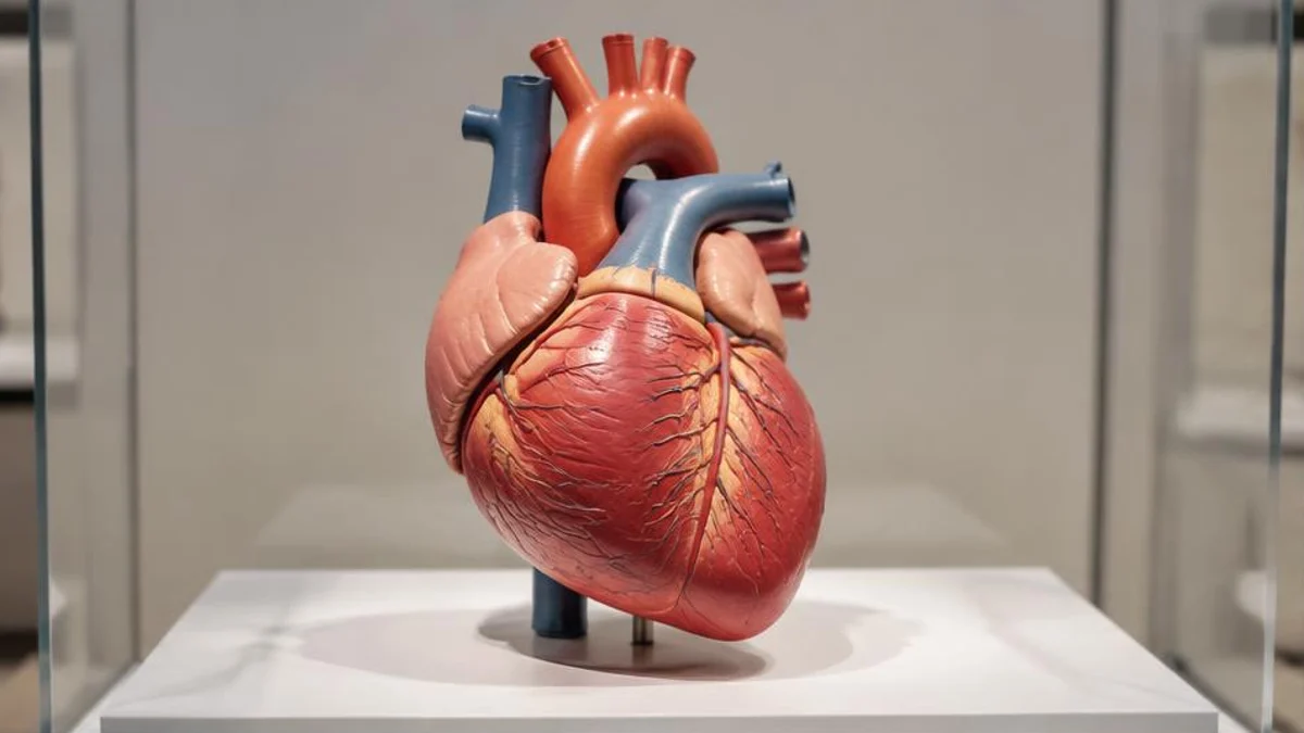
The heart sits slightly left of the centre of your chest and is divided into a right half and a left half by a muscular wall. Each half has two chambers:
- Atria (upper chambers): the right atrium and left atrium receive blood returning to the heart.
- Ventricles (lower chambers): the right ventricle and left ventricle push blood out of the heart.
The ventricles have thicker walls than the atria because they do more work. The left ventricle has the thickest wall of all, since it must push blood through the whole body, while the right ventricle only needs to push blood through the nearby lungs.
Two circuits, one heart
Blood travels through two loops that join end to end.
The pulmonary circuit (to the lungs)
Blood that has already delivered oxygen to the body returns to the heart through two large veins, the superior vena cava and the inferior vena cava. It enters the right atrium, passes through the tricuspid valve into the right ventricle, and is then pushed through the pulmonary valve into the pulmonary arteries. These arteries lead to the lungs, where blood picks up oxygen and releases carbon dioxide.
The systemic circuit (to the body)
Oxygen-rich blood returns from the lungs through the pulmonary veins into the left atrium. It then passes through the mitral valve into the left ventricle. When the left ventricle contracts, it pushes blood through the aortic valve into the aorta, the body's largest artery. From there, branching arteries deliver blood to the brain, muscles, digestive system and every other tissue. After the tissues take what they need, the blood flows back through veins to the vena cavae, and the cycle begins again.
The two circuits operate at the same time. While the right side sends blood to the lungs, the left side sends blood to the body, and every beat does both.
Arteries and veins: a common mix-up
Many people learn that arteries carry oxygen-rich blood and veins carry oxygen-poor blood. That is true for most of the body, but it is not the definition. The definition is about direction:
- Arteries carry blood away from the heart.
- Veins carry blood toward the heart.
The pulmonary circuit shows why this matters. The pulmonary arteries carry oxygen-poor blood away from the heart to the lungs, and the pulmonary veins carry oxygen-rich blood from the lungs back to the heart. Using direction as the rule avoids confusion.
What the valves actually do
The heart has four valves, and they work like one-way doors. It is tempting to imagine them being opened and closed deliberately, but they are passive structures. Each valve is made of thin flaps of tissue that are pushed open when pressure behind them is higher than pressure in front, and pushed closed when the pressure reverses.
- The tricuspid and mitral valves sit between the atria and ventricles.
- The pulmonary and aortic valves sit where blood leaves the ventricles.
When a ventricle contracts and pressure rises, the valve between the atrium and ventricle snaps shut so blood cannot flow backward, and the exit valve opens so blood can leave. When the ventricle relaxes, the pressure drops, the exit valve closes, and the entry valve opens again.
The sound of a heartbeat
The familiar "lub-dub" sound of a heartbeat comes largely from valves closing. The first sound ("lub") is made when the tricuspid and mitral valves close at the start of ventricular contraction. The second sound ("dub") is made when the aortic and pulmonary valves close as the ventricles relax. Clinicians listen to these sounds with a stethoscope because unusual sounds can sometimes signal a problem, though only a professional assessment can say what a particular sound means.
What keeps the beat steady
Heart muscle cells can generate and conduct electrical signals. A small group of specialised cells in the right atrium, called the sinoatrial (SA) node, acts as the heart's natural pacemaker. It fires a signal at a regular rate, and that signal spreads across the atria, causing them to contract. The signal then passes through a relay point and travels down into the ventricles, which contract a fraction of a second later.
That delay is important. It gives the atria time to finish pushing blood into the ventricles before the ventricles squeeze. The nervous system and hormones can speed up or slow down the SA node, which is why your heart rate rises when you exercise or feel anxious and drops when you rest.
Systole and diastole
One heartbeat has two main phases:
- Diastole: the heart relaxes and the chambers fill with blood.
- Systole: the heart contracts and pushes blood out.
The chambers do not all contract at once. The atria contract first to top up the ventricles, then the ventricles contract to send blood out. This coordination makes the heart an efficient pump.
When blood pressure is measured, the two numbers reflect these phases. The higher number (systolic) is the pressure in the arteries when the heart contracts, and the lower number (diastolic) is the pressure when the heart relaxes between beats.
The heart also needs its own blood supply
Blood passing through the chambers does not feed the heart muscle directly. The heart has its own network of vessels called the coronary arteries, which branch off the aorta and wrap around the surface of the heart. They deliver oxygen and nutrients to the heart muscle itself. Because the heart never rests, a steady supply through these vessels is essential.
Putting it together: one trip around the loop
Here is the whole route in order, starting with blood returning from the body:
- Blood enters the right atrium through the vena cavae.
- It passes through the tricuspid valve into the right ventricle.
- The right ventricle pushes it through the pulmonary valve to the lungs.
- In the lungs, the blood picks up oxygen.
- It returns through the pulmonary veins to the left atrium.
- It passes through the mitral valve into the left ventricle.
- The left ventricle pushes it through the aortic valve into the aorta.
- Arteries deliver it to the body, and veins return it to the start.
Frequently asked questions
Does blood only travel in one direction through the heart?
Yes, in a healthy heart. The valves prevent backflow, so blood moves forward through the chambers in a fixed order.
Why does the left side of the heart have thicker walls?
The left ventricle pushes blood through the entire body, which requires more force than sending blood to the nearby lungs. Thicker muscle lets it generate that pressure.
Do the atria and ventricles contract at the same time?
No. The atria contract slightly before the ventricles. This brief delay lets the ventricles fill completely before they pump.
Is the heart's electrical signal the same as an ECG?
An electrocardiogram (ECG) records the heart's electrical activity from the surface of the body. It is a tool for measuring the signals described above, and interpreting it takes clinical training.
Sources and further reading
Related reading on FactNest: How vaccines train the immune system and 10 fascinating facts about the human brain.

Written by
FactNest Editorial Team
FactNest is operated by Muonix. The FactNest Editorial Team publishes general-interest explainers with AI-assisted drafting and illustrations. Contact contact@muonix.co for questions or corrections; we respond within 2 business days.
More from this author →